Carotid
CEA
Unusual carotid enlargement after CEA
An 80-year-old patient with coronary disease, diabetes, and a history of stroke presents with a left carotid patch enlargement and tight stenosis before the venous patch—yet the carotid bifurcation and distal internal carotid remain patent.
With a complex vascular history, including previous carotid surgery and recurrent strokes, this case raises a critical question: How would you manage the next steps?

Bordeaux, France

Hospital Pellegrin - Bordeaux, France
Professor of vascular surgery
Professor of vascular surgery
Head of unit of vascular surgery CHU Bordeaux - France
Conflicts of interest:
Honoraria from: COOK Médical, Térumo-Vascutek, Siemens, GORE
Part I - Case presentation
A 80-year-old patient presented with :
- Coronary diseases / Diabetic status / Dyslipidemia
- Previous carotid surgical treatment on left side with venous patch closure in 2014 in another peripheral center
- Previous history of stroke in 2016 and 2022 with excellent recovery. Clinical transient history of right brachial deficit and paresthesia
- Recent investigation with Angio CT scan showing:
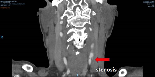
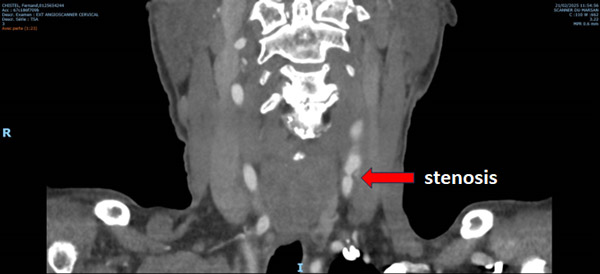
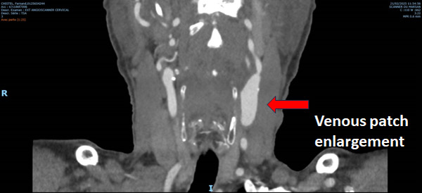
- Left carotid patch enlargement without thrombus
- Left common carotid tight stenosis before venous path
- Carotid bifurcation patent
- Distal left internal carotid patent without stenosis
Part II - Our treatment
Our decision:
- Right common femoral artery access
- Catheterism of left common carotid
- Embolisation of external carotid artery
- Exclusion on carotid enlargement with self-expanding covered stent
- Angioplasty of proximal stenosis
- Dual anti-platelet medication post operative period
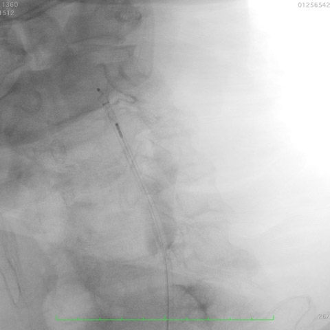
Per operative angiogram from right femoral access
- Carotid bifurcation enlargement
- Tight stenosis at the proximal edge of the venous patch
- External carotid artery patent
- Absence of lesion on the internal carotid artery
Per operative embolisation of external carotid artery
- Direct catheterism with 0.018 wire
- Direct 7 Fr sheath placement
- Angiogram control
External carotid embolisation
- 5 Fr plug placement at the ostium of external carotid artery to avoid any retrograde perfusion
- Control with perfect plug placement and provisional arterial perfusion maintained
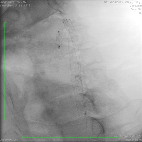
Exclusion of carotid enlargement
- 0.018 guidewire placement in internal carotid artery
- No protection device added
- Viabahn 7 x 75 mm delivered
- Precise deployment and delivery
Final angioplasty
- Proximal angioplasty on tight stenosis at 8 atm using 6 x 4 mm balloon
- Distal angioplasty at 4 atm same balloon
- Angiogram control showing excellent result without residual stenosis
Intra-cerebral angiogram
- Absence of embolism
- Perfect perfusion without defect
- Patient delivered at D+ 1
- Dual anti-platelet medication during at least 3 months
Get the latest clinical cases and breaking news delivered straight to your inbox!
